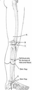
With advances in the field of prostheses is the selection of the amputation in order to maintain the limb sedistal may not be entirely correct. This applies to the superior limb amputation. Rules which states for limb mempretahankan sedistal may not be applicable to the inferior extremity amputation. Even so far as possible the knee must be saved, because the knee is very useful functionally. Problem of weight bearing and leaves soft tissue to cover the stump greatly influence the selection of the amputation of the inferior ekstremias. In below knee amputation stump that is too long is not advised because it would complicate the use of the prosthesis. Anterior border of the tibia should be available in the bevel and enough soft tissue to cover it by making a flap diposterior longer. Amputation of ankle-high enough indication has rarely, usually in trauma. Syme amputation is useful for end weight bearing prosthesis. For amputation of the foot is general agreement that is used is trans metatarsal (the level of amputation see schematic drawing).
Location to perform an amputation:


b. Operation indication
* Trauma
* Dead ganggan limb due to vascular supply
* Malignant neoplasms
* Chronic Osteomyelitis
* Life-threatening infections
* Congenital limb deformities are inoperable
c. Contra indications of operation: the general state of poor
Engineering Operations
Management of Extremity Amputation
Anesthesia
Spinal anesthesia is commonly used for lower extremity amputations, anstesia common for upper limb amputation. Can also be used leksus block anesthesia. Amputation of the finger can be used for local infiltration anesthesia.
Mechanical operation
Above-knee amputation
The best place to split the femur is 8-10 cm (the width of one hand). Use of skin markers to plan the incision, which should create a flap of anterior and posterior flaps have the same length or slightly longer anteriorly. For those of skin and subcutaneous tissue along the line are planned. Hemostasis is usually not difficult in the ischemic limb but severe bleeding can occur in the limbs are septic. Tie all of veins by using absorbent needle 2 / 0. The anterior incision is deepened to the bone, cutting the quadriceps femoris tendon. Vasa femoral together media and lateral popliteal nerves found in posteromedial position. Tie with thread veins double absorbency. Before cutting the nerve, give stress on the nerve so nerve interested in the amputation stump. If the amputation performed at a higher level, sciaticus nerve can be found. Sciaticus nerve followed by the artery to be didiseksi separately and fastened prior to nerve cut. After cutting all the muscles around the femur, the living tissue vessels and avoid the use of diathermy. Check the exact point of amputation of the femur and scrape the periosteum from the bone in this area. The muscles of the thighs must be retracted to the proximal direction to provide sufficient space in the use of chainsaws. This can be done with the help of some abdominal pads or special retractors. After cutting off the femur and lower leg, place a clean towel under the butt and rest your butt on the inverted bowl. Use stingy to smooth the edge of the femur, then bring the muscles along the front and back cover cut off the bone with suture thread absorbency size 1. Attach suction drain the skin incision below the point of cutting the bone in the muscle layer. Place the second layer of stitches is more superficial in the muscle and subcutaneous tissue as this will help close the flap of skin. Sew the edge of the skin with stitches broke up with non-absorbent yarn 2 / 0. Avoid picking the edge of the skin with toothed forceps. Close stump with gauze and cotton and dressing with crepe bandage.
Below-knee amputation
Optimum point for amputation is 14 cm from the tibial plateau, the fibula was cut 2 cm proximal from this. Tick incision, with anterior flap ends just distal of the cutting line on the tibia bone and the posterior flap extends down to the Achilles tendon. Make an incision along the lines that have been marked. In the posterior Achilles tendon cut and deepen the incision to cut the rest of the muscles and tendons to bone. Cut across the muscle into up front. Fibular oblique cut with a saw Gigli, then split the tibia 2 cm distal to this. Clean the muscle from the bone with periosteum elevator. Cut the anterior bevel first with a saw and cut perpendicular to the diagonal of the tibia. Forms angle at the lower end of the tibia towards the top and separate the muscle mass of the posterior aspect. Tie a double all the blood vessels and cut every nerve tense. Remove the distal limb. The posterior flap is pulled upward to wrap butt bone and sutured to the anterior flap. Posterior flap may need to be reduced by excision of muscle tissue. Place absorbent yarn in between the muscles in the posterior and anterior subcutaneous tissue and leave the suction drain beneath the muscle. Bring the edge of the skin with stitches drop out of non-absorbent yarn 2 / 0. Snip the corners of the posterior flap if necessary in order to form neat. Close the butt with a tight bandage with cotton and crepe bandage.
Complications of surgery
* Bleeding
* Infection
Mortality
Depending on the etiology
Postoperative care and follow-up
* Wound care in general
* Rehabilitation of the manufacture of a suitable prosthesis
* Rehabilitation of the manufacture of a suitable prosthesis
No comments:
Post a Comment